At the intersection of art and science is the annual Nikon “Small World in Motion” video competition, offering a glimpse into the microscopic world unseen to the human eye — with a little assistance from microscopic technology, that is.
And this year, the five winners of the annual contest bring us one glance deeper into the tiny elements and organisms that our world has to offer.
“Nikon’s Small World is regarded as the leading forum for showcasing the beauty and complexity of life as seen through the light microscope,” wrote Nikon on its homepage.
The video competition encompasses movie or digital time-lapse photography taken through the microscope.
Fifth Place: Showcasing the World's Deadliest Animal
Mosquitoes have been dubbed by health experts as the “world’s deadliest animal” because of the diseases they spread — dengue, yellow fever, lymphatic filariasis, to name a few — killing more people than any other animal in the world. Though their reach is mighty, they are small. A video captured by Dr. Sachie Kanatani of Johns Hopkins Bloomberg School of Public Health and Dr. Photini Sinnis of the Department of Molecular Microbiology and Immunology showed an infected mosquito salivating fluorescently marked malaria parasites.
The fifth-place winner was captured using what is known as confocal imagery, a technique that scans a specimen to create computer-generated optical sections via visible light. These sections are then stacked to provide a 3-dimensional reconstruction of the specimen at hand.
Fourth Place: Mapping the Nervous System
Neurons make up a complex internal map of living organisms, and Dr. Alexandre Dumoulin of the University of Zurich Department of Molecular Life Sciences captured just that in his fourth-place entry. The video showed commissural axons “turning into an organized manner” after having crossed the midline of the central nervous system.
A midline is the central feature in bilateral organisms and holds within it highly specialized cells. For example, the spinal system in humans makes up our midline. And contained within the midline of some organisms are a type of neuron known as commissural axons that help signal various sensations to organisms. As is seen in this video, commissural axons grow and spread in a somewhat chaotic fashion before changing directions and growing longitudinally toward the brain, according to University College London.
Third Place: The Birthing of a Water Flea
Captured by microscopic photographer Andrei Savitsky from Ukraine, the third-place winning video shows a water flea, scientific name Daphnia pulex, giving birth to several cubs.
According to the biodiversity database Animal Diversity Web, D. pulex is a common species of water flea and is found in almost every type of freshwater — from “rain-filled tire ruts” to tree moss in a rainforest. These tiny organisms are similar to their land flea cousin yet are light enough to stay suspended in the water column, using their legs and antennae for movement.
As can be seen in the video, their folded, shell-like surrounding structures, called a carapace, is clear and allows a viewer to see most of the flea’s internal organs and heart. A dark-colored eye can also be seen through the insect’s clear head.
The video was captured using a technique known as darkfield that creates contrast in transparent specimens, allowing their bodies to shine through a central light.
Second Place: Watching an Engineered Tumor Form
A 10-day time-lapse captured by Dr. Stephanie Hachey and Dr. Christopher Hughes of the University California, Irvine Department of Molecular biology and Biochemistry shows an engineered human micro-tumor as it forms and metastasizes. The vessels, which are shown in red, support the growth of the tumor highlighted in blue.
Using confocal and fluorescence, a high-intensity illumination to “excite fluorescent molecules” in a sample, the researchers were able to showcase how tumors can spread through the human body.
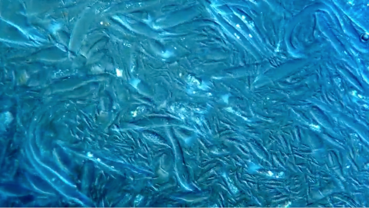
First Place: The Symbiotic Relationship of Termites and Protists
First place was awarded to Fabien Weston of Sydney, Australia, whose close-up video shows microfauna in a termite gut.
Microorganisms found in the guts of termite, also known as protists, play an essential role in digesting plant-based cellulose such as wood. These single-celled organisms form a symbiotic relationship with termites to process cellulose, help the bugs to derive nutrition from it, and cycle carbon back into the soil.
“Protists, while largely unknown to the general public, are indeed the most abundant creatures on the planet,” said Weston. “There is a significant gap in our understanding about these termite symbionts and how this unique evolutionary relationship developed with its host, making it well worth exploring and presenting."
The blue-hued video was captured using polarized light passed through a filter in a research microscope from the 1970s. Weston created an environment with a pH, chemical composition and temperature that would keep the symbionts alive.
“The most challenging part of capturing this video was finding the right solution for the creatures themselves,” said Weston. “I tried a lot of methods, even preparing my own saline solution. They’re very sensitive to oxygen, so I had to remove as much gas from the solution as possible. It was very tricky, and I had to work fast. The video you’re seeing is the result of months of trial and error, a lot of research and perseverance.”
Honorable Mentions
Can’t get enough of the world behind a microscope? From mudflat diatoms to pooping worms and a flailing sea cucumber, you can check out the dozens of honorable mentions here.