
This degree of magnification and resolution is made possible by the use of a Focused Ion Beam Scanning Electron Microscope, or FIB-SEM. Ordinary microscopes will not produce the same results.
Curious about how Snopes' writers verify information and craft their stories for public consumption? We've collected some posts that help explain how we do what we do. Happy reading and let us know what else you might be interested in knowing.
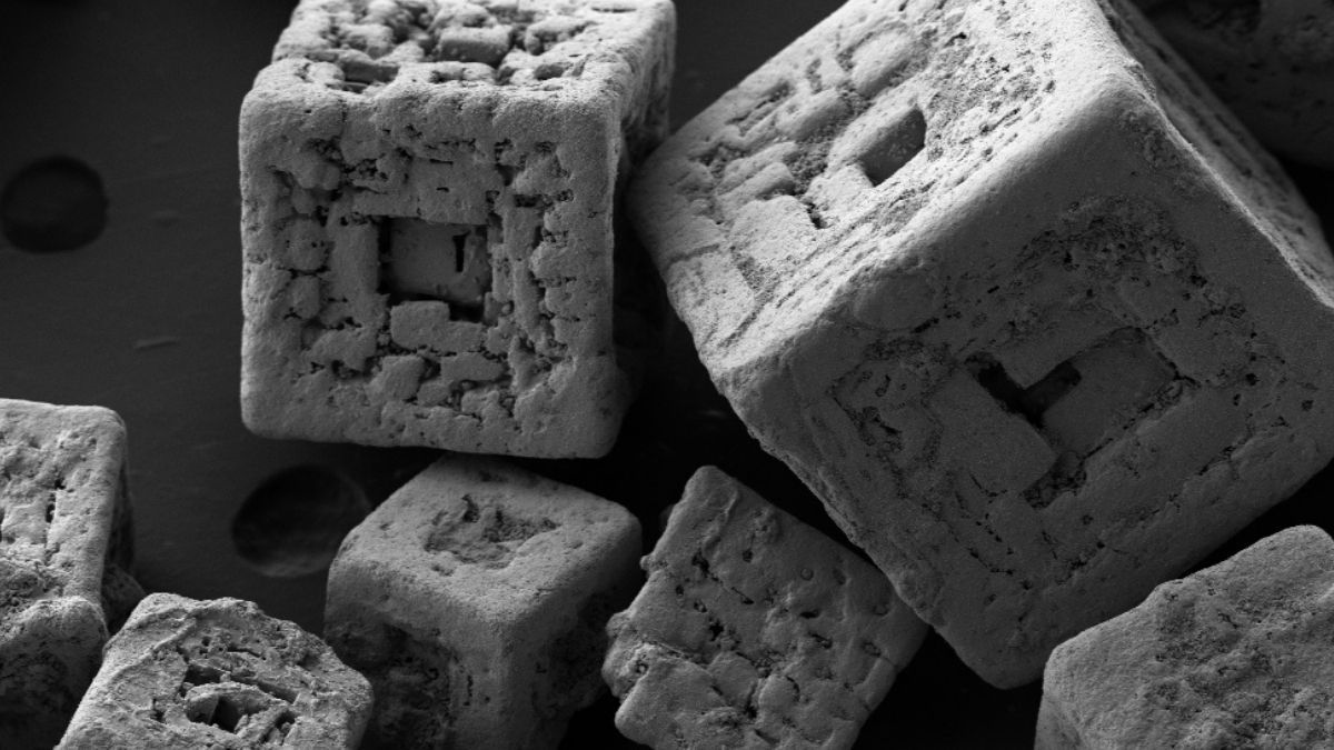
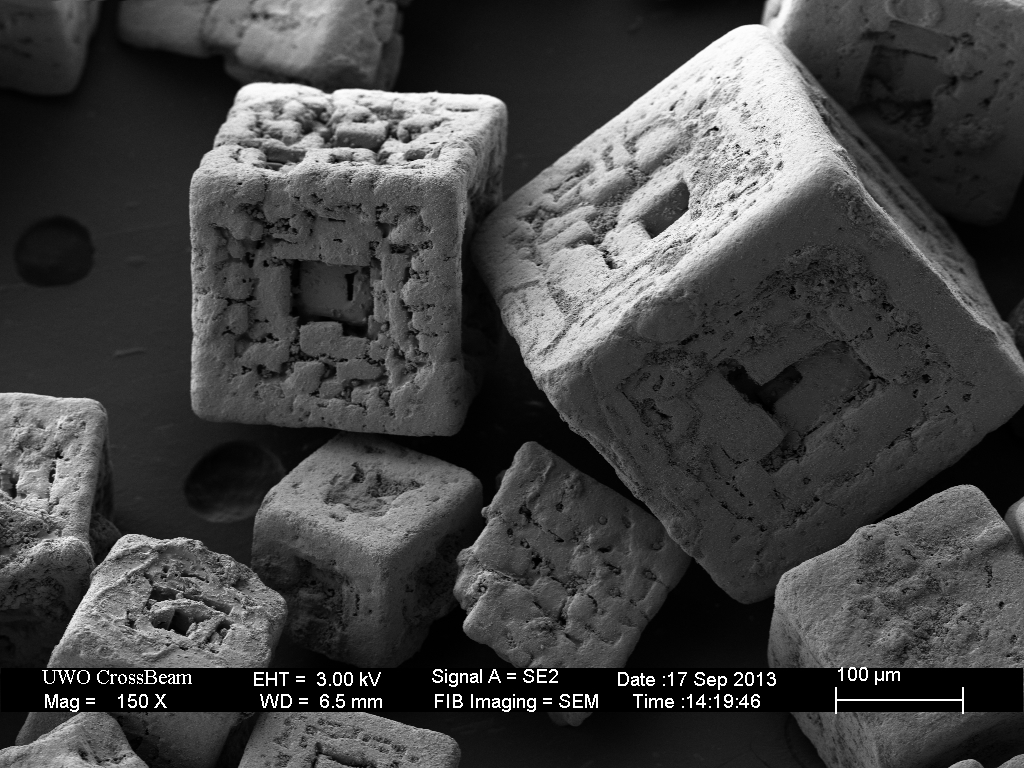
An image supposedly showing "Grains of salt under an electron microscope" is frequently met with skepticism after it gets shared on social media:
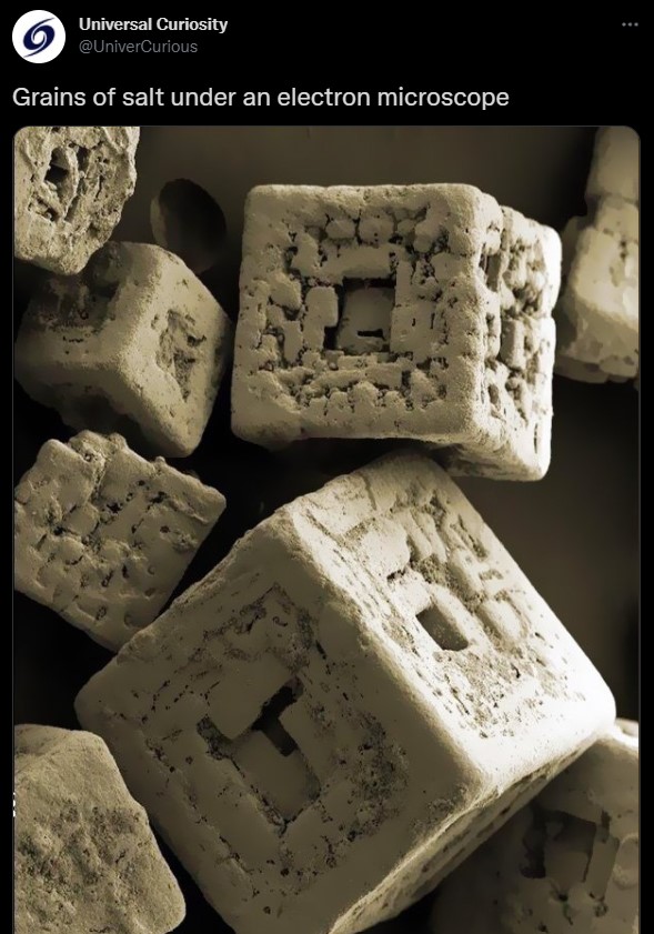
While these large blocks may not resemble table salt to the naked eye, this is a genuine image of grains of salt under a microscope.
This image was taken at the Nanofabrication Facility at Western University in Ontario, Canada. It shows "fine salt" (french fry salt) at 150x magnification. It was originally posted on Flickr by ZEISS Microscopy in 2014 along with the following caption:
Fine salt, 150x
Crystals of fine salt, imaged with 150x in the FIB-SEM. Kindly provided by Todd Simpson, UWO Nanofabrication Facility
The image can also be found on UWO Nanofabrication Facility's Tumblr page:
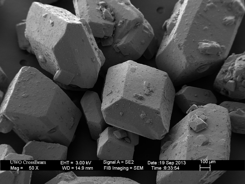
Here's a similar image that was taken at UWO Nanofabrication Facility. Instead of fine salt, this image shows sugar:
While these images genuinely show salt and sugar under a microscope, don't expect to get the same results with your home or school equipment. The images were produced with a high-powered (and expensive) scientific instrument called a FIB-SEM, or Focused Ion Beam Scanning Electron Microscope, that uses a focused beam of ions to scan and create 3D images.
A paper published by eLifeSciences in 2017 explained: "Focused Ion Beam Scanning Electron Microscopy (FIB-SEM) can automatically generate 3D images with superior z-axis resolution, yielding data that needs minimal image registration and related post-processing."
Here's a video explaining more about the FIB-SEM: