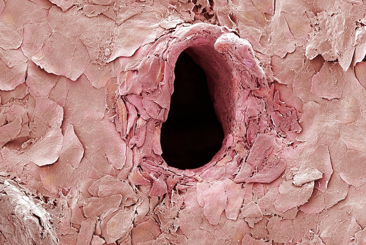

An image shared to Reddit on April 11, 2021, claimed to depict an intimate look at a puncture hole in skin made by a needle — and its flaky, almost cave-like attributes garnered the picture more than 22,800 upvotes on the social media platform.
A reverse image search revealed that the photograph in question was hosted by Science Photo Library, a London-based database of science and medical images. The SPL website noted that its images are "acquired from scientific and medical experts, acclaimed photographers and renowned institutions."
According to the caption, the photo was captured by Anne Weston of Francis Crick Institute and showed an up-close-and-personal, highly magnified puncture in the skin made by a tattoo needle:
Tattoo needle puncture in skin, coloured scanning electron micrograph (SEM). Skin tattooing has been practised for thousands of years. To make tattooing permanent tiny needles are used to punch through the top layer of the skin (epidermis) and into the next layer (dermis). This image shows many skin cells from the epidermis surrounding a hole created by a tattoo needle. Magnification: x280 when printed at 10cm wide.
Tattoos become permanent by piercing the top layer of the skin and depositing a small amount of ink within each puncture, according to the Mayo Clinic. And that microscopic process was captured and magnified by SEM, a photography process that the National Library of Medicine noted allows for the "topographical visualization of structures."
An SEM is more powerful than a traditional microscope because the device scans an electron beam over an item, rather than using a combination of light and high-power lenses to amplify a subject. These electron beams interact with a given sample to map out its surface topography. It works in a similar way as some other, more large-scale topographical mapping technologies, such as deep-sea mapping of the seafloor or light detection and ranging (LiDAR) processes on terrestrial landscapes — but at a much smaller scale.
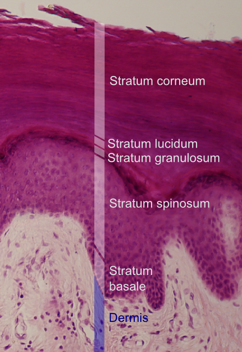
The SEM picture shows the top layer of the skin known as the epidermis. Its outermost layer is known as the stratum corneum, Latin for "horny layer." This outer layer provides the first line of defense for the body against the external world and is made up of corneocytes. Corneocytes are made up of keratin filaments — flat cells that organize in a "brick and mortar formation."